Spectroelectrochemistry is a multi-response technique that provides electrochemical and spectroscopic information about a chemical system in a single experiment, i.e., it offers information from two different points of view. Raman spectroelectrochemistry could be considered as one of the best techniques for both the characterization and behavioral understanding of carbon nanotube films, as it has traditionally been used to obtain information about their oxidation-reduction processes as well as the vibrational structure. This application note describes how the SPELECRAMAN is used to characterize single-walled carbon nanotubes by studying their electrochemical doping in aqueous solution as well as to evaluate their defect density.
Raman spectroelectrochemistry is one of the most powerful techniques for the characterization of carbon nanomaterials. Carbon nanotubes (CNTs) in particular are one of the most interesting materials to study via electrochemical purposes due to their exceptional mechanical, electronic, and thermal properties. Raman spectroelectrochemistry could be considered as one of the best techniques for both the characterization and behavioral understanding of CNT films. This multi-response technique has traditionally been used to obtain information about their oxidation-reduction processes as well as the vibrational structure of CNT films.
The SPELEC RAMAN with a 785 nm laser is used to characterize single-walled carbon nanotubes (110SWCNT electrode) as well as to study their electrochemical doping in aqueous solution. In this Application Note, the anodic charging was studied by scanning the potential from 0.00 V to different upper potentials and back to 0.00 V at 0.05 V s-1 scan rate in 0.1 M KCl aqueous solution. Raman spectra were recorded using an integration time of 1 s, allowing dynamic analyses to be performed.
Characterization of 110SWCNT was performed by Raman spectroscopy. The Raman spectrum of SWCNT (Figure 1) exhibits four major bands: radial breathing mode (RBM), disorder induced mode (D), tangential displacement mode (G) and high frequency two phonon mode (G’).

RBM is detected between 120–300 cm-1 and provides suitable information about the diameter of the nanotubes. There are as many different CNT diameters in the sample as there are RBM bands differentiated in the spectrum. The relationship between the RBM frequency, ωRBM (cm-1), and the diameter of the CNT, dt (nm), is given by the following equation:
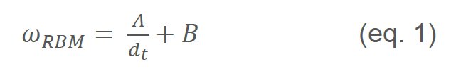
where A (nm cm-1) and B (cm-1) are semiempirical parameters comprising of values between 220–230 nm cm-1 for A and 10–20 cm-1 for B, depending on the experimental conditions [1]. As shown in the inset of Figure 1, four RBM bands are differentiated in 110SWCNT. According to eq. 1 and taking into account the position of RBM bands (Figure 1, inset), the calculated diameters are 1.55 nm, 1.19 nm, 1.07 nm, and 0.92 nm.
Furthermore, electrochemical doping of 110SWCNT was studied by analyzing the spectroelectrochemical behavior of the G-band. This band is associated with the tangential vibration modes of the nanotubes and provides information not only related to the metallic or semi-metallic character of the nanotubes but also about the doping process.
Figures 2a and 2b show Raman spectra recorded from 0.00 V to +1.00 V during the forward and backward scans, respectively. Evolution of Raman intensity of the G-band (at 1592 cm-1) with potential is represented in Figure 2c. As can be observed, the G-band decreases from 0.00 V to +1.00 V and the minimum intensity is reached at +1.00 V. In the reverse scan, Raman intensity recovers an almost similar intensity to the initial value at the end of the experiment. The position of the G-band is not modified during the electrochemical doping. Changes in Raman intensity are related to the depletion/filling of Van Hove singularities in the SWCNT electronic density states, and G-band bleaching is associated with changes in the resonance condition [2].
 and reverse (b) scan. (c) Evolution of the G-band with potential during the anodic charging process.")
Electrochemical p-doping was also studied from 0.00 V to more positive potentials. Figure 3 shows the Raman spectra obtained by scanning the potential from 0.00 V to +1.80 V. Figure 3a shows how the G-band decreases in the forward scan. However, in the reverse scan (Figure 3b), Raman intensity increases only slightly and it does not approach the values of the initial intensity.
Furthermore, not only is the bleaching of this band observed, but also an up-shifting during the anodic charging (Figure 3c). This effect on the position of the G-band is explained by changes in the spring force constant of C-C bonds, and also by the phonon renormalization [3].
 and reverse (b) scan. (c) Spectroelectrochemical behavior of the G-band during the anodic charging process.")
Time-resolved Raman spectroelectrochemistry also provides valuable quantitative information. As the G-band intensity does not depend on defects, the ID/IG intensity ratio has been traditionally used for the evaluation of the defect density. Table 1 shows the ID/IG ratio at different positive potentials, demonstrating that the ratio increases with potential. Comparison between ID/IG at +1.00 V and +1.80 V shows that the ratio is more than 2 times higher at +1.80 V than at +1.00 V because more defects are electrochemically generated in the SWCNT film.
| Upper potential | ID/IG intensity ratio |
|---|---|
| + 1.00 V | 0.51 |
| + 1.20 V | 0.80 |
| + 1.40 V | 0.91 |
| + 1.60 V | 1.16 |
| + 1.80 V | 1.26 |
Raman spectroscopy is one of the best methods to characterize SWCNT due to the resonant enhancement of the Raman signal. Furthermore, time-resolved Raman spectroelectrochemistry is a powerful technique to study dynamic chemical systems, like the charging process of carbon nanotubes. Evolution of Raman intensity of the Gband with potential increases understanding about the characterization of SWCNTs. Finally, the combination of Raman spectroscopy and electrochemistry provides suitable information to evaluate the defect density of a SWCNT structure.
- M.S. Dresselhaus, G. Dresselhaus, R. Saito, A. Jorio, Raman spectroscopy of carbon nanotubes, Physics Reports. 409 (2005) 47–99.
- L. Kavan, L. Dunsch, Spectroelectrochemistry of carbon nanostructures., ChemPhysChem. 8 (2007) 974–998.
- M. Kalbac, L. Kavan, L. Dunsch, Effect of Bundling on the Tangential Displacement Mode in the Raman Spectra of Semiconducting Single- Walled Carbon Nanotubes during Electrochemical Charging, J. Phys. Chem. C. 113 (2009) 1340–1345.
 Share via email
 Download PDF